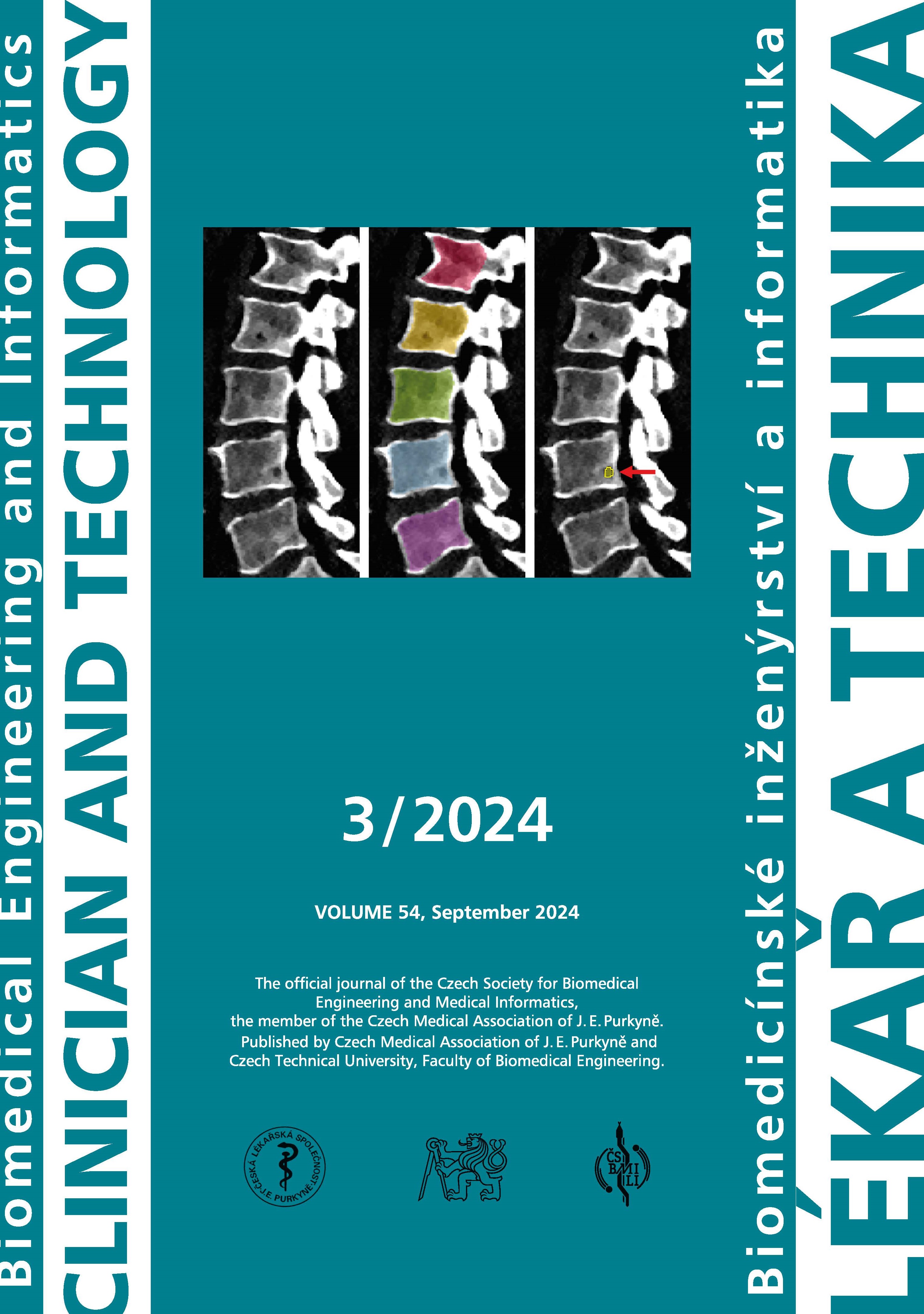
METHOD COMPARISON FOR BONE DENSITY IN MULTIPLE MYELOMA PATIENTS
DOI:
https://doi.org/10.14311/CTJ.2024.3.04Abstract
Bone mineral density (BMD) is an important indicator of bone health, particularly in patients with conditions such as multiple myeloma. This study aims to compare three methodologies for quantifying BMD in vertebral regions affected by lytic lesions: two using data from conventional CT with different corrections for tissue composition, and one using data acquired on a dual-energy CT system. Method 1 is based on conventional CT with corrections using reference values for muscle and fat, Method 2 uses conventional CT with corrections based on the measured CT values of paraspinal muscle, and Method 3 is based on dual-energy CT. The Wilcoxon signed-rank test was used for statistical comparison, as the dataset did not follow a normal distribution. The results indicated significant differences between Methods 1 and 2 for BMD in regions of interest (ROIs) within lytic lesions, while no significant differences were found for other comparisons in this group. For vertebrae affected by multiple myeloma, significant differences were found between Methods 1 and 2, and Methods 2 and 3, but not between Methods 1 and 3. In healthy vertebrae, a significant difference was found only between Methods 2 and 3. When all ROIs were combined, significant differences were found between Methods 1 and 2, and Methods 2 and 3, with no difference between Methods 1 and 3. Future research will focus on objectively assessing the accuracy of these methods by comparing their results with a calibration phantom.
Downloads
Published
Issue
Section
License
Copyright (c) 2024 Michal Nohel, Martin Mezl, Vlastimil Valek, Marek Dostal, Jiri Chmelik

This work is licensed under a Creative Commons Attribution 4.0 International License.
Authors who publish with this journal agree to the following terms:
- Authors retain copyright and grant the journal right of the first publication with the work simultaneously licensed under a Creative Commons Attribution License (https://creativecommons.org/licenses/by/4.0/) that allows others to share the work with an acknowledgment of the work's authorship and initial publication in CTJ.
- Authors are able to enter into separate, additional contractual arrangements for the non-exclusive distribution of the journal’s published version of the work (e.g., post it to an institutional repository or publish it in a book), with an acknowledgment of its initial publication in this journal.
- Authors are permitted and encouraged to post their work online (e.g., in institutional repositories or on their website or ResearchGate) prior to and during the submission process, as it can lead to productive exchanges.
CTJ requires that all of the content of the manuscript has been created by its respective authors or that permission to use a copyrighted material has been obtained by the authors before submitting the manuscript to CTJ. CTJ requires that authors have not used any copyrighted material illegally, as for example a picture from another journal or book, a photo, etc. It is the author’s responsibility to use only materials not violating the copyright law. When in doubt, CTJ may ask the authors to supply the pertinent permission or agreement about the use of a copyrighted material.
The opinions expressed in CTJ articles are those of authors and do not necessarily reflect the views of the publishers or the Czech Society for Biomedical Engineering and Medical Informatics.


